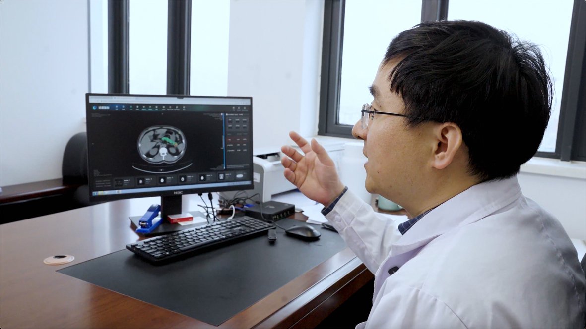
The decisive moment in pancreatic-cancer AI is not the colored region on a scan. It is the clinical handoff that follows. In March 2025, a Chinese report described a 65-year-old patient who went to Ningbo University Affiliated People's Hospital because of dizziness. Routine examinations had not raised a pancreatic alarm, but an AI review of his non-contrast CT flagged a lesion reported as 1.5 centimeters. Further examinations confirmed the finding, and he underwent surgery. By that point, the hospital said it had used the system to examine more than 40,000 people and found eight pancreatic cancers, six at an early stage.[3]
The system was PANDA, short for pancreatic cancer detection with artificial intelligence, developed by researchers from Alibaba's DAMO Academy and collaborating medical institutions.[1][2] A January 2026 Xinhua report said the Ningbo deployment had expanded beyond 180,000 abdominal and chest CT scans since late 2024, uncovering more than 20 pancreatic cancers, including 14 early-stage cases.[4] Those reported outcomes are promising. They also reveal the right way to judge the use case: not by how many scans an algorithm can process, but by whether an alert survives radiologist review, reaches the patient, earns an appropriate confirmatory test, and changes care without creating an unmanageable trail of false alarms.
As of July 17, 2026, PANDA is best understood as an unusually well-studied screening assistant moving through clinical trials and live Chinese hospital workflows—not as an autonomous diagnosis or a settled case for population-wide pancreatic-cancer screening.[1][3][4][5]
The useful input is a scan that already exists
PANDA's strongest product idea is opportunistic screening. It does not ask a healthy population to undergo a new CT examination solely for pancreatic cancer. It re-reads a non-contrast chest or abdominal CT that was acquired for another clinical reason: a checkup, an emergency visit, a lung examination, or an unrelated symptom. Running software on that existing image adds no radiation exposure by itself. Any later contrast CT, MRI, endoscopic ultrasound, biopsy, or surgery is a separate decision that must be justified by the alert and the patient's clinical picture.[1][5]
That distinction matters. The U.S. Preventive Services Task Force's current final recommendation, issued in 2019, advises against screening asymptomatic average-risk adults for pancreatic cancer. Its evidence review found no proof that general-population screening improved morbidity or mortality and emphasized the harms that can follow false positives and invasive workups.[5] That recommendation does not adjudicate PANDA's later evidence or govern Chinese practice. It does establish the problem PANDA must solve: finding a rare, deadly cancer early is valuable only if the detection method is specific enough, and the downstream pathway disciplined enough, that benefit can outrun harm.
The 2023 Nature Medicine paper explains how the model tries to extract more from the borrowed scan. PANDA first localizes the pancreas with an nnU-Net, then uses convolutional networks to detect a possible lesion, then classifies the finding across pancreatic ductal adenocarcinoma and seven non-PDAC categories with help from a memory-transformer branch. Its supervision came from pathology-confirmed labels and lesion outlines transferred from paired contrast-enhanced CT, where abnormalities are easier to see, onto the corresponding non-contrast images.[1] It is therefore neither a generic vision model nor an image generator. It outputs a lesion boundary and probabilities learned from deliberately paired clinical evidence.
The reproducibility boundary is also material. The paper says the implementation code cannot be released because it depends on internal infrastructure and is patent-protected. Its competing-interest statement identifies Alibaba patent applications related to the method and says several authors were Alibaba employees who owned company stock through their compensation.[1] Those disclosures do not negate peer review; they make independent prospective validation more important.
A strong retrospective study is still retrospective
The research program is substantial. PANDA was trained on 3,208 patients at one high-volume Shanghai center, then tested across external centers and imaging protocols. The paper reports a 5,337-patient external multicenter cohort, a separate 492-patient chest-CT cohort, and two consecutive real-world cohorts totaling 20,530 patients.[1] The associated Chinese trial registration describes the work as a retrospective diagnostic study of opportunistic screening on non-contrast CT, an important boundary for interpreting the result.[1]
The chest-CT experiment illustrates both the opportunity and the danger. In that selected cohort, 67% of patients with PDAC did not have the entire lesion captured in the chest scan's field of view. PANDA could sometimes respond to secondary signs such as a dilated pancreatic duct, but a partial view cannot become a reliable all-clear. The model may notice a reason to look again; it cannot make anatomy outside the image visible.[1]
The real-world retrospective cohorts provide the more useful operational numbers. In the first 16,420-patient cohort, PANDA identified 26 pancreatic or nearby lesions that the initial standard-of-care assessment had not detected. Eight were subsequently found through ordinary follow-up before the retrospective AI study occurred. Of the remaining patients invited for MRI during the COVID-19 period, only one complied; surgery confirmed a 1.5-centimeter grade 1 pancreatic neuroendocrine tumor.[1] Across both retrospective cohorts, the paper says PANDA found five cancers and 26 other pancreatic lesions missed at the initial visit.[1]
That is meaningful evidence of signal. It is not evidence that 31 AI alerts independently produced 31 clinical rescues. Timing, follow-up, and patient participation sit between detection and benefit. The paper itself respects that boundary: it calls for prospective studies of risk, benefit, and cost-effectiveness and defines PANDA as a screening step before diagnosis, not a replacement for established diagnostic imaging.[1]
The base rate moves the burden downstream
Specificity is the pivotal number because pancreatic cancer is uncommon. Under the paper's initial lesion-detection definition, PANDA produced 156 apparent false-positive alerts in the first 16,420-patient cohort, corresponding to a raw specificity of 99.0%. A multidisciplinary team judged 80 of those alerts to be other pancreatic or nearby diseases that still merited radiologist attention. After that reclassification, 76 patients—0.5%—remained false positives, 70 of which radiologists could readily rule out; the adjusted specificity was 99.5%.[1]
The word adjusted deserves attention. Clinically, some of the 80 reclassified findings may be valuable. Operationally, they still consume review time and can still lead to a callback. A hospital needs both views: whether the model was wrong about its target, and whether the alert created useful work, unnecessary work, or both. The team subsequently used hard-example mining and incremental learning to produce PANDA Plus. On a separate 4,110-patient cohort, the upgraded model reduced false positives by more than 80% while retaining sensitivity, reaching an adjusted specificity of 99.9% for lesion detection and PDAC identification.[1]
Even 99.9% specificity does not abolish the base-rate problem. Using a general-population PDAC prevalence of 13 per 100,000 adults, the authors estimated that PANDA could produce roughly 11 true positives and 100 false positives per 100,000 tests—a positive predictive value near 10%.[1] That projection may still compare favorably with established screening programs, but it means most positive alerts in a very low-prevalence population could be false. The callback pathway is therefore not administrative plumbing. It is part of the clinical intervention.
The live Ningbo reports make the missing denominator visible. The public record tells us that more than 180,000 scans yielded more than 20 pancreatic cancers, 14 reportedly early-stage.[4] It does not yet disclose, in a peer-reviewed prospective analysis, how many total alerts were generated, how many radiologists dismissed, how many patients were recalled, which confirmatory tests they received, how many invasive procedures proved unnecessary, or how many cancers the system missed. Without those counts, case stories show that the pathway can work; they do not yet measure its full tradeoff.
The callback loop is the deployment unit
A defensible PANDA deployment should be audited as a sequence: eligible existing scan, model version, alert threshold, radiologist decision, patient contact, confirmatory imaging, multidisciplinary judgment, pathology when obtained, treatment, and follow-up. Each step needs a denominator and a timestamp. Hospitals should report false negatives as carefully as celebrated detections, separate PDAC from other lesions, and preserve the distinction between an AI flag, a radiologic suspicion, and a confirmed diagnosis.[1][5]
Model evolution belongs in that ledger too. PANDA Plus improved after learning from hard examples and false positives encountered outside the original training center.[1] That is a reasonable engineering response, but every update changes the device being evaluated. Performance should be tracked by model version, hospital, scanner protocol, patient group, and time period so that improvement at a familiar site does not hide drift elsewhere. The 2023 paper also notes that its data outside East Asian populations and hospitals were limited and that pancreatic neuroendocrine tumors remained a weaker category.[1]
The forward test is concrete. Prospective multicenter evidence should show that the system maintains performance on consecutive patients, keeps the callback burden tolerable, shifts cancers toward resectable stages, and closes follow-up quickly enough for earlier detection to alter outcomes. The falsifier is equally concrete: if scan volume and success stories grow while hospitals cannot publish alert denominators, missed-cancer audits, unnecessary-workup rates, and patient outcomes, then PANDA remains a promising detector inside an unmeasured care pathway.
One next-stage study is already registered. The ClinicalTrials.gov record for PANDAPro, updated June 5, 2026, lists an estimated enrollment of 100,000 and an active, not recruiting status. Its prospective round is designed to record model results in real time, recall discordant cases, and follow apparent false positives for as long as two years. No results are posted, and overall completion is estimated for August 2027.[6] The enrollment target is therefore a statement of intended scale, not a performance result.
The Ningbo photograph captures the right division of labor. A doctor sits between the model's highlighted region and the patient's next step.[3] PANDA's achievement is to make a quiet part of an ordinary scan worth a second look. Its proof will come from what the health system does with that look.
Sources
- Kai Cao et al., “Large-scale pancreatic cancer detection via non-contrast CT and deep learning,” Nature Medicine 29 (2023)—model design, reader studies, multicenter and chest-CT validation, retrospective cohorts, false-positive analysis, limitations, code availability, competing interests, and clinical-use boundary.
- DAMO Medical AI, “Latest Research in Nature Medicine: CT Achieves Large-scale Pancreatic Cancer Screening for the First Time Based on DAMO Academy's Medical AI” (November 2023)—developer account of the collaboration, paired-image training logic, architecture, and intended opportunistic-screening use.
- Economic Information Daily via Sina Technology, “AI precisely breaks the deadlock in pancreatic-cancer screening” (March 21, 2025)—Ningbo clinical-trial workflow, early deployment figures, case details, and source page for the documentary photograph.
- Xinhua, “AI in China spots cancers doctors might have missed” (January 14, 2026)—reported Ningbo deployment scale, early-stage case count, patient follow-up, and expansion to county facilities.
- U.S. Preventive Services Task Force, “Final Recommendation Statement: Pancreatic Cancer: Screening” (August 6, 2019)—general-population screening recommendation, evidence gaps, low-prevalence effects, and downstream harms.
- ClinicalTrials.gov, “Research of the Application of Pancreatic Cancer Screening Artificial Intelligence Model ‘PANDAPro’” (NCT06643715; record updated June 5, 2026)—study status, estimated enrollment, retrospective and prospective rounds, recall workflow, follow-up plan, and estimated completion.